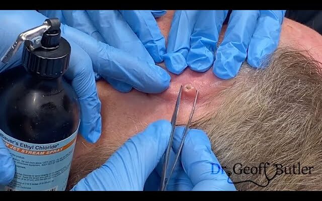
The removal of squamous cell carcinoma (SCC) from behind the ear typically involves a surgical procedure to remove the cancerous tissue and ensure clean margins, which means taking out a small amount of healthy tissue around the cancer to reduce the risk of recurrence. Squamous cell carcinoma is a type of skin cancer that is often caused by prolonged sun exposure, but it can also occur in other areas of the skin or mucous membranes.
Here’s an overview of what to expect if you’re having SCC removed from behind your ear:
Steps for Removal:
- Initial Consultation: A dermatologist or a surgeon will evaluate the tumor and assess the best course of action. They may perform a biopsy to confirm that the growth is indeed squamous cell carcinoma and determine its stage and depth.
- Anesthesia: For this procedure, local anesthesia is typically used. The area around the SCC will be numbed so that you won’t feel pain during the procedure. In some cases, especially for larger or more complex tumors, general anesthesia may be recommended.
- Incision: The surgeon will make an incision around the tumor, taking care to remove not just the cancerous tissue but also a small border of healthy tissue to ensure that no cancer cells remain. The location behind the ear may involve careful planning to minimize scarring, as this area is visible and often sensitive.
- Excision: The SCC and surrounding tissue are removed, and the specimen is sent to a lab for pathology to confirm clear margins (i.e., no cancerous cells left behind).
- Wound Closure: After the tumor is removed, the surgeon will close the incision. In most cases, sutures are used, and depending on the size of the incision, it may be necessary to use layered stitches (deeper layers and then surface layers) to close the wound securely.
- Post-Procedure Care: After the procedure, you will be given instructions for care, which typically include:
- Keeping the area clean and dry
- Applying any prescribed ointments (like antibiotics) to reduce the risk of infection
- Watching for signs of infection, such as increased redness, swelling, or drainage
- Avoiding sun exposure to the area while it heals
- Follow-Up: A follow-up appointment is usually scheduled to check on the healing process and to remove the stitches if non-dissolvable sutures were used. If the excised tissue is confirmed to have clear margins, no further treatment may be necessary. However, if cancer cells were found at the edges of the removed tissue, additional treatment (such as Mohs surgery, radiation, or further excision) may be required.
Risks and Considerations:
- Scarring: Since the procedure is near the ear, there may be visible scarring, though skilled surgeons can minimize scarring with precise techniques.
- Infection: As with any surgical procedure, there’s a risk of infection at the incision site, so it’s important to follow aftercare instructions.
- Recurrence: Although SCC is usually treatable with surgery, there is a possibility of recurrence, especially if the margins were not clear or if the cancer was aggressive.
- Facial Nerve Damage: Given the proximity to the facial nerve and the ear, special care is taken during the procedure to avoid damaging surrounding structures.
Prognosis:
- Squamous cell carcinoma, especially when caught early, has a high cure rate with surgical removal. The prognosis is generally excellent for people with localized SCC that hasn’t spread to lymph nodes or distant areas of the body.
After removal, you’ll likely be monitored for any signs of recurrence, and your doctor may recommend regular skin checks as part of your ongoing care plan, especially if your SCC was caused by sun exposure.
If you’re going through or considering this treatment, make sure to talk through any concerns with your doctor, as they can provide personalized advice based on the specifics of your case.


